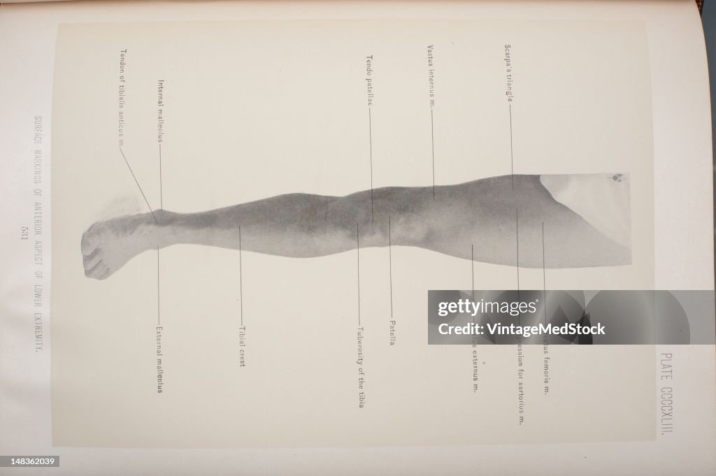
Surface Markings Of Anterior Aspect Of Lower Extremity
Illustration from 'Surgical Anatomy: The Treatise of the Human Anatomy and Its Applications to the Practice of Medicine and Surgery, volume III' (by Dr. John Blair Deaver) shows Scarpa's triangle, rectus femoris, depression for satorius, vastus externus, patella, tuberosity of the tibia, tibial crest, external malleolus, tendon of tibialis anticus, internal malleolus, tendo patellac, vastus internus, 1903. (Photo by VintageMedStock/Getty Images)

PURCHASE A LICENCE
How can I use this image?
£275.00
GBP
Getty ImagesSurface Markings Of Anterior Aspect Of Lower Extremity, News Photo Surface Markings Of Anterior Aspect Of Lower Extremity Get premium, high resolution news photos at Getty ImagesProduct #:148362039
 Surface Markings Of Anterior Aspect Of Lower Extremity Get premium, high resolution news photos at Getty ImagesProduct #:148362039
£375£150
Getty Images
In stockPlease note: images depicting historical events may contain themes, or have descriptions, that do not reflect current understanding. They are provided in a historical context. Learn more.
DETAILS
Restrictions:
Contact your local office for all commercial or promotional uses.
Credit:
Editorial #:
148362039
Collection:
Archive Photos
Date created:
01 January, 1903
Upload date:
Licence type:
Release info:
Not released. More information
Source:
Archive Photos
Object name:
T1674624_190
Max file size:
4256 x 2832 px (36.03 x 23.98 cm) - 300 dpi - 4 MB