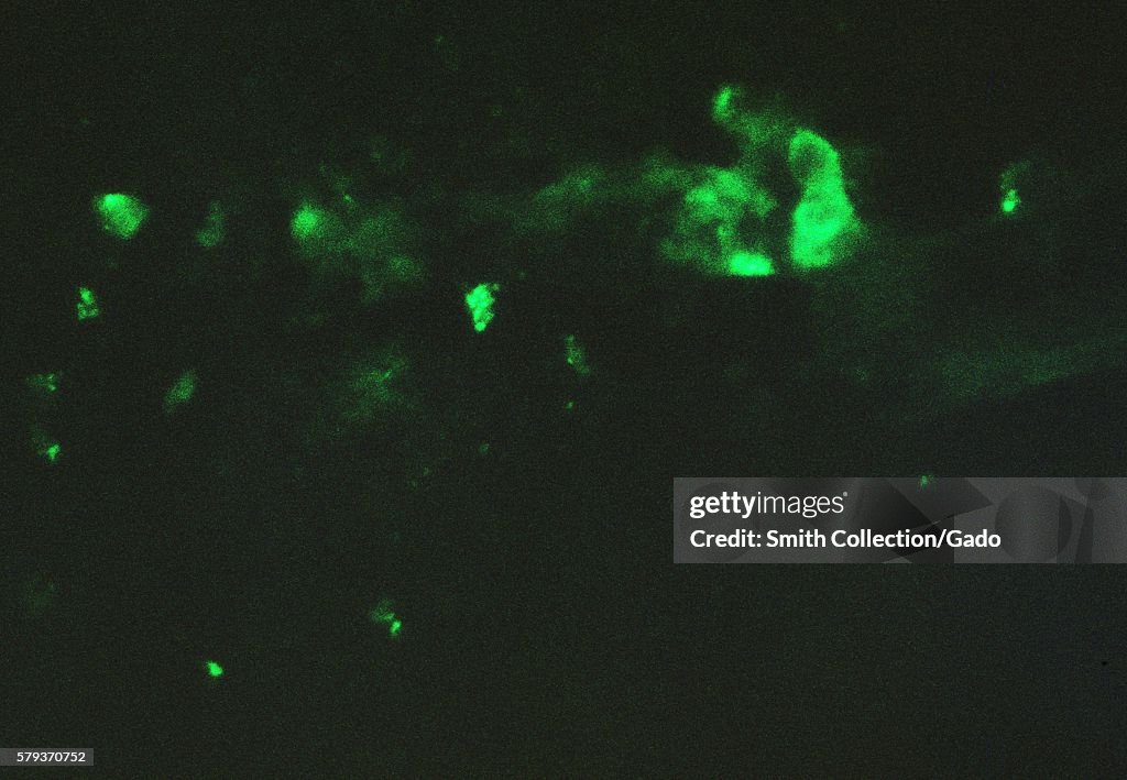
Chick Chorioallantoic Membrane
A photomicrograph of a chick chorioallantoic membrane, fluorescent antibody staining was done after smallpox virus inoculation, 1962. Chorioallantoic membrane infected with variola, stained with rabbit anti-vaccinia conjugate, then viewed using immunofluorescent microscopy technique. Image courtesy CDC/Dr. David Kirsh. (Photo by Smith Collection/Gado/Getty Images).

PURCHASE A LICENCE
How can I use this image?
£375.00
GBP
Please note: images depicting historical events may contain themes, or have descriptions, that do not reflect current understanding. They are provided in a historical context. Learn more.
DETAILS
Restrictions:
Contact your local office for all commercial or promotional uses.
Credit:
Editorial #:
579370752
Collection:
Archive Photos
Date created:
01 January, 1962
Upload date:
Licence type:
Release info:
Not released. More information
Source:
Archive Photos
Object name:
77951final.jpg
Max file size:
5400 x 3741 px (45.72 x 31.67 cm) - 300 dpi - 5 MB